

Thirty years after Arwen Bird was rear-ended and paralyzed by a speeding drunk driver, she started hearing ringing sounds in her head. “I decided to have imaging done because I wanted to understand what caused the ringing, and in particular why I was hearing my heartbeat at the same time,” says Bird, now a T8 para.
CT imaging showed both carotid arteries had aneurysms — arterial bulges — which can cause ringing in the ears, also known as tinnitus. Her doctor theorized that the aneurysms in her neck were likely due to the C7/T1 dislocation she sustained in addition to the T6-8 burst fracture. He decided it was best to repeat imaging at least every three years — to guard against unexpected clotting or arterial bleeding or rupturing, any of which could cause serious damage or even death. He also prescribed a daily low-dose of baby aspirin to lessen the risk.
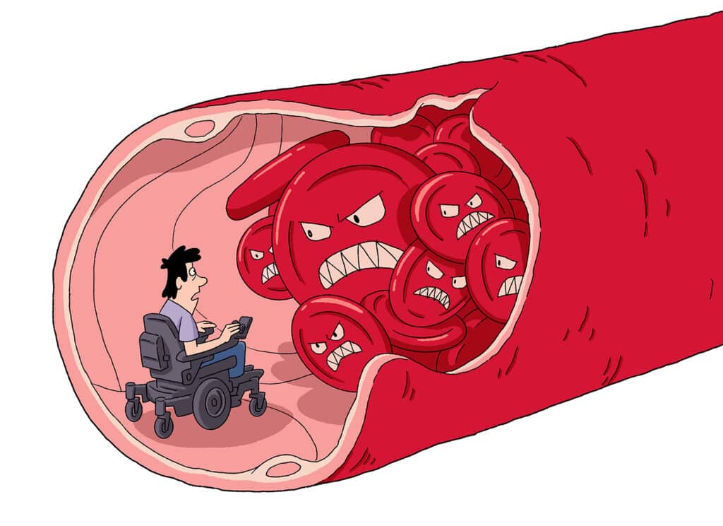
Bird’s discovery of her aneurysms three decades after her accident serves as a warning to all of us. By not knowing what’s going on inside our bodies, which most of us do not since we have no way of routinely checking our arterial health, we are at risk every day. While clotting is a danger, at a deeper level this story is also about the health of our arteries — the mostly invisible freeway system that carries life-giving blood to every part of our bodies.
The first three months following SCI are the period of greatest downtime in bed and the least activity, which makes clotting more likely.
With traumatic spinal cord injury, the danger of clotting, especially in the lower extremities, begins immediately. Dr. Joseph Shatzel is a hematologist and scientific researcher at Oregon Health & Science University who has won multiple teaching awards and has published numerous research papers and book chapters on hematological topics. Among his colleagues, his expertise has earned him the nickname Dr. Clot, and on X he goes by @clotmaster.
He says the greatest risk of clotting for an individual with SCI comes immediately following the injury. “When someone is just injured — probably during the first three months — they are at a very high risk for … blood clots that occur in the veins of the legs,” he says. “Sometimes they can migrate into the lungs.” A clot in the lungs is known as a pulmonary embolism.
The Lowdown on Blood Clots and Blood Thinners
Not all blood thinners are the same. There are about 18 blood thinners on the market. All treat clotting, but they work in different ways. The two main groupings are anticoagulants and antiplatelet medications. The former, more commonly used, slow down the making of platelets in the first place, while the latter group prevents platelets from binding together to form clots and are usually prescribed for those who have had a heart attack or stroke or are at high risk of having one. Peripheral artery disease, which is usually caused by plaques that narrow lower extremity arteries and reduce blood flow, can also require antiplatelet treatment. For more on clotting and blood thinners, visit:
• UW Medicine, “Blood Clot Risk in People with SCI”
• Healthline.com, “Everything You Need to Know About Deep Vein Thrombosis”
• MedlinePlus, “All About Blood Thinners”
Embolisms and Blood Thinners
I didn’t have to look far to find someone in our community who had personal experience with early clotting and a life-threatening pulmonary embolism. Bob Vogel, 65, who I’ve worked with at New Mobility for the last 25 years, developed a dangerous clot after a 1985 skiing accident that broke four ribs and crushed his T11-12 vertebrae. “It was massive trauma,” says Vogel. “They did a spinal fusion and installed Harrington rods within a week. About two weeks after that, I was just lying there, way doped up, still groggy, talking to my dad, and I just mentioned a sharp pain, not all that bad, whenever I breathed in.” A nurse came in and listened to him breathe through a stethoscope, followed by a doctor who did the same. “The next thing I know I’m being wheeled down a hallway for an emergency operation.”
They found a pulmonary embolism in his right lung and traced the origin to his right thigh. Vogel was put on Lovenox, an anticoagulant, right away, then Coumadin, a different anticoagulant, for another six months until clots in his lung and right thigh dissolved. “I was good to go,” says Vogel.
Then in 2007 he flipped forward out of his chair and broke his right femur near where he had the clot in his thigh. He and his doctor noticed the swelling wasn’t going down after the fracture. An ultrasound showed another clot had formed, so once again doctors put him on Lovenox, then Coumadin. About five months later, a follow-up showed the clot in his thigh had not completely dissolved, but the blood flow had found a way around it. Nonetheless, with his doctor’s approval, Vogel decided to stay on Coumadin indefinitely. “Doctors told me once you’ve had a clot, your chances of having another increase,” he says.
Finally, in 2018, after not having clots for more than 10 years, he decided it was safe to discontinue blood thinners. He only had clotting problems following traumatic injuries. He remains vigilant, keeping close watch on any possible swelling. He also has written down his history of clotting and taking blood thinners and keeps a copy of it in his wallet, in case it is ever needed. “Knock on wood,” he says, “no problems since then.”
What Research Tells Us
A 2015 study conducted at the National Spinal Injuries Center at Stoke Mandeville Hospital in the U.K. found that even though blood thinners are routinely prescribed, in the first 90-day period of SCI, the rate of PE ranges from 5-12%. On the other hand, the incidence of PE was only 1.25% in patients who were more than 90 days post-SCI (and no longer taking blood thinners). The takeaway is clear: The chance of getting a clot following onset of SCI is highest during the first few months even with blood thinners, but it drops off rapidly after that. In the general population, the rate of PE is 0.06% per year.
It should be noted that the first three months following SCI are the period of greatest downtime in bed and the least activity, which makes clotting more likely. There is also a higher rate of clotting with quads, but it is not all that different for those with thoracic or lumbar injuries. Everyone is at risk, but the risk is relatively small, and blood thinners are always an option to reduce the risk significantly.
Recurring Danger
Teal Sherer, 44, another NM associate, was injured at L2 in a 1995 car accident in Knoxville, Tennessee. A few weeks after lengthy hospital and rehab stays, she was back in school as a 14-year-old freshman, when a blood clot came out of the blue. “I woke up one morning and my left leg was swollen,” she says. “We’d been warned of blood clot danger, so right away I knew what was happening.”
She went to the hospital and thinks she was given a blood thinner via IV and sent home after several days. She’s still foggy on the details. “But I remember when I was discharged, I was definitely not on a blood thinner or at least not for that long,” she says. She stayed active, walking around the house with long leg braces, moving her legs, swimming in outpatient rehab. It was all good, no more blood clots.
Fast-forward to 2014. Married and pregnant, her maternal fetal doc put her on low-dose Lovenox twice daily, because she was at higher risk of forming new clots because of her blood clot history. “He told me, ‘You had a clot before, now you’re pregnant for the first time, let’s just do this and be safe,’” she says.
A year after giving birth to her son, River, she fell out of her chair and broke an ankle. Two weeks later she got another blood clot in her left leg. (Clots are more likely to form after a traumatic accident.) “I had to go to the ER with a swollen leg, then was sent home with blood thinners,” she says.
“After this, I found out I had a compressed vein in my left hip, [known as] May-Thurner syndrome, which is more common in women who have given birth,” says Sherer. “MTS restricts return blood flow and is fairly common in women 20 to 40 who have a blood clot history.”
After moving to Seattle, to be safe, she sought out a vascular doctor, who decided to put a stent in her compressed vein and open it up. The stent collapsed. A second try at stenting was successful. “It’s all good now. I have to wear a compression sock and I take Eliquis (another blood thinner),” she says. “Because of my history, I’m on top of my health now. I have my doctors, and I see them regularly. I’m aware of potential problems, even when doctors sometimes aren’t. They should have put me on blood thinners when I broke my ankle. Now I know, so I’m thinking and being proactive.”

What About Carotid Arteries?
If you have a chronic SCI and wonder if your carotid arteries might have been damaged from a traumatic accident, like Bird, you can always request a carotid artery ultrasound scan. It is a common noninvasive preventative procedure that can easily be arranged. If the ultrasound study finds you are at risk from arterial damage or narrowing from plaque buildup, chances are that stenting or a routine procedure to remove plaque can be done. If an aneurysm is discovered, further testing will be done to assess the degree of risk and whether it is best to operate. Sometimes the best option is to do nothing — or possibly take a daily baby aspirin or some other blood thinner.
How many of us might be at risk of arterial problems? In the case of car crashes, the percentage of SCI cases with carotid artery damage is not known, but assuming it mirrors that of the mainstream population, it could be in the 1-2% range. On the other hand, since traumatic vehicular accidents are the leading cause of SCI, the percentage could very well be higher. No matter the cause or incidence, it is important for all SCI survivors to be aware of potential arterial damage from clots or narrowing and take preventative action, if necessary.
Support New MobilityWait! Before you wander off to other parts of the internet, please consider supporting New Mobility. For more than three decades, New Mobility has published groundbreaking content for active wheelchair users. We share practical advice from wheelchair users across the country, review life-changing technology and demand equity in healthcare, travel and all facets of life. But none of this is cheap, easy or profitable. Your support helps us give wheelchair users the resources to build a fulfilling life. |
